A Simple Mole Check
Can Save Your Life
Skin And Cancer Institute founder and board-certified dermatologist and skin cancer surgeon Dr. Daniel Taheri highly recommends his patients to undergo routine skin examinations to prevent and detect skin cancer or any other dangerous growths such as moles or pre-cancerous skin lesions. He also advises against excessive sun exposure and strongly supports wearing high SPF protection to avoid a diagnosis of skin cancer.
What causes skin cancer?
Skin cancer is caused by the uncontrolled replication of mutated cells in the skin. There are several major categories of skin cancer, including Squamous Cell Carcinoma, Basal Cell Carcinoma and Melanoma. Each type develops from a specific class of mutated cells. Less common types of skin cancer include Merkel Cell Carcinoma, Sebaceous Gland Carcinoma and Kaposi Sarcoma.
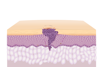
Squamous Cell Carcinoma
Squamous cells that mutate and replicate create tumors consisting of crusty, scaly, or bloody patches and sometimes elevated lesions or warts.
Signs and Syndroms
Squamous cell carcinoma can develop in people of any skin type, but it is prevalent in individuals with fair skin. A squamous cell carcinoma typically starts out as a lesion with a crusty or scaly surface or as a firm, red nodule. In fair-skinned individuals, the carcinoma usually develops on the hands, face, ears, and other sun-exposed areas of the body. In people with darker skin, the nodules or lesions may develop in areas that are not often exposed to sunlight.
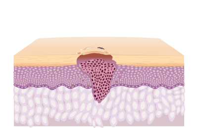
Basal Cell Carcinoma
Basal Cell Carcinoma is generated when basal cells replicate uncontrollably deep inside the epidermis, forming tumors that look like open sores, patchy growths, oozing bumps, or scars.
Signs and Syndroms
Basal cell carcinoma is the most commonly diagnosed form of skin cancer. It usually occurs on the face, neck, or other sun-exposed areas of the body and is most often seen in people with fair skin. The carcinoma may present as a brown or flesh-colored lesion or as a bump that appears pearly or waxy.
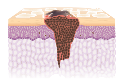
Melanoma
Melanoma is caused by a mutation in pigmented skin cells, known as melanin.
Signs and Syndroms
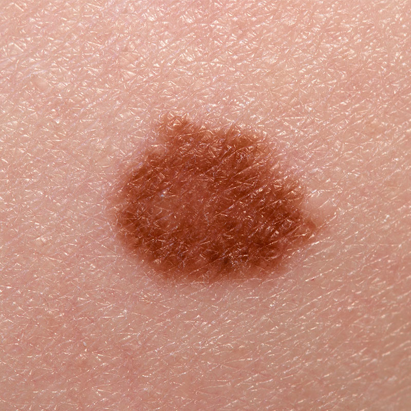
Melanoma is an aggressive and potentially deadly form of skin malignancy. The carcinoma often develops in a mole or as a dark spot on the skin. These malignancies typically develop on the trunk or face in males and on the lower legs in females. It is possible for melanoma to develop in areas that are not normally exposed to the sun, including the palms, soles of the feet, and under the fingernails and toenails of individuals with dark skin. It is also possible for the malignancy to affect the mucous membranes of the nose, mouth, anus, or vagina.
Mohs Micrographic Surgery
Skin cancer comes in various forms, with basal cell carcinoma and squamous cell carcinoma being the most prevalent. Melanoma and Merkel cell carcinoma are rarer types of skin cancer. The size, location and type of lesion on the body or skin is an indication of the type of skin cancer present and typically lets the surgeon or dermatologist make a diagnosis.
Mohs micrographic surgery is a special technique used by skin cancer plastic surgeons to eliminate skin cancer from the skin and to remove tumors from the face. This technique removes cancerous tissue layer by layer. Each layer is inspected and analyzed by a lab to ensure that no cancerous cells remain in the tissue. Mohs surgery is the preferred skin cancer removal method by many surgeons because it allows for the maximum preservation of healthy non-cancerous tissue and skin which is important to our patients.
The Mohs Step-by-Step Process
This skin cancer treatment involves removing any layers of skin that contains cancer cells. The procedure starts with the removal of a small area of skin cancer, which is immediately tested under a microscope onsite to determine the location of the cancer cells in the sample. Patients remain in the operating room during testing. Once analysis pinpoints the direction of the cancer’s growth, another small layer of skin is removed and examined. This process continues layer by layer until all skin cancer cells have been removed.
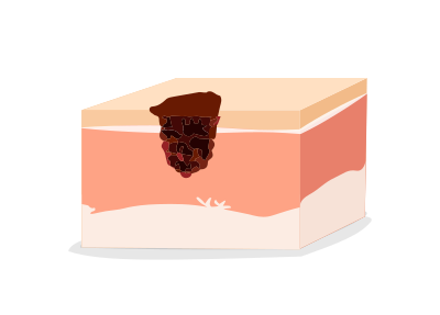
Step 1
Under local anesthesia, the patient undergoes removal surgery by scalpel as portions of the tumor are excised by the dermatologic surgeon.
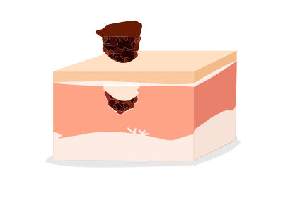
Step 2
All of the cancerous tissue is eventually yet slowly excised by the board-certified dermatologist and skin cancer surgeon.
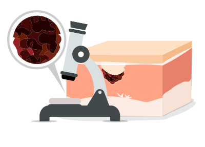
Step 3
The tumor and tissue are sent to a lab and analyzed. This is sometimes enough to remove the cancer entirely.

Step 4
If the lab results show the cancer was completely removed, the procedure is complete. If the lab results show remaining cancerous tissue, the process repeats itself and the surgeon removes another layer of skin.
Types of tumors and skin cancer include basal cell, squamous cell, merkel cell, melanoma and sebaceous carcinoma.
Screening for moles is critical because a mole that changes in size, color or appearance can become cancerous.
If a skin lesion changes in texture, size, color, itches or begins to ooze or bleed, the precancerous lesion has turned into skin cancer.
Mohs surgery is a highly effective skin cancer removal procedure that is increasingly popular especially when the tumor is on the face.
Patient Testimonials
Explore our patient portal of testimonials regarding their experience with skin cancer removal, their treatment and specifically the Mohs surgery technique with Dr. Daniel Taheri.
Patient Testimonials
Explore our patient portal of testimonials regarding their experience with skin cancer removal, their treatment and specifically the Mohs surgery technique with Dr. Daniel Taheri.
Will I have a Scar After Mohs Surgery?
Scarring may occur as a result of the Mohs surgery; however, it will be minimized to the extent possible by your surgeon. If you follow the post-surgery wound instructions from your doctor, you can further minimize the appearance of any scars.
Patient #1
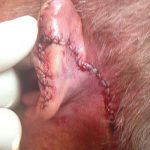
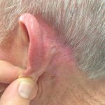
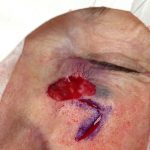
Removal of skin cancer behind left ear.
Patient #2
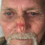
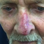
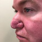
Removal of skin cancer on the nose.
Patient #3
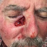
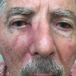
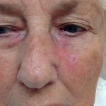
Removal of skin cancer below the right eye.
Patient #4
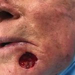
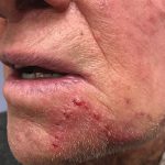
Removal of skin cancer on left side of chin.
Patient #5
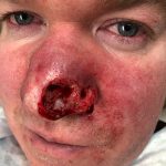
Patient #6
Why are skin cancer screenings and biopsies important?
Full body cancer screenings and biopsies are the first line of defense against skin cancer. Regular cancer screenings ensure that abnormal growths on the skin are detected before they progress into more dangerous forms, and biopsies ensure that the growths are accurately diagnosed.
What happens during a full body skin cancer screening?
Patients who receive a skin cancer screening at Skin And Cancer Institute can expect a quick and painless 10-15 minute exam. A skin cancer specialist will check the entire body for atypical moles and unusual lesions, paying close attention to size, color, border, and shape. If anything looks suspicious, a biopsy will be ordered.
What happens when a biopsy is taken?
Biopsies are samples of body tissue taken in order to test for cancer cells and other harmful conditions. Sometimes a biopsy will be small, and other times a doctor may need to use stitches to close the skin surrounding the biopsied area. Some patients experience mild discomfort when undergoing a biopsy procedure, which is why local anesthesia is used before the tissue sample is excised. Once testing is complete, a specialist will contact patients to discuss biopsy test results and next steps for treatment.
Biopsy Methods
The two most common biopsy methods include the shave and the punch techniques. Your doctor will select the best method for your biopsy based upon the location and type of skin abnormality.
Shave Biopsy
The shave biopsy method is used when a lesion raises above the surface of the skin. A scalpel is held parallel the lesion and then moved in a straight motion to remove the skin sample.
Punch Biopsy
The punch biopsy is advantageous in flat lesions where the shave technique would leave behind a visible divot in the skin. For this method, the entire lesion is removed if cancer is likely present. When cancer is unlikely, only the darkest, most elevated area is removed. Using a specialized punch tool selected to fit the exact lesion size, the instrument is pressed downward with a back and forth turning motion. Forceps are then applied to elevate the skin sample, and scissors are used to snip the skin from fatty tissue. Punch biopsies require stitches to close the wound.
How many types of skin cancer are there? Does each type of skin cancer look different?
Atypical Moles are unusual looking moles that are either noncancerous or precancerous. The more atypical moles a patient has, the more likely they are to develop skin cancer. When multiple atypical moles are present, a biopsy is usually taken to determine whether the moles are cancerous. Click here to learn more about abnormal moles.
Melanoma is a type of skin cancer that can develop from common and atypical moles as well as healthy skin. It is caused by a mutation in pigmented skin cells, known as melanin. This melanin is produced and delivered throughout the skin by cells called melanocytes. Sometimes excess melanin and melanocytes cluster together, causing moles. Within these clusters, melanocytes may mutate or trigger surrounding cells to mutate, causing skin cancer. Click here to learn more about melanoma.
Actinic Keratoses are precancerous crusty, scaly clusters within the epidermal skin layer that can mutate into cancer. Most often, actinic keratosis spots turn into squamous cell carcinoma if left untreated. Click here to learn more about actinic keratosis.
Squamous Cell Carcinoma The epidermal layer of skin is made of squamous cells. When these cells mutate and replicate uncontrollably, they form tumors that present as crusty, scaly, or bloody patches and sometimes elevated lesions or warts. Click here to learn more about squamous cell carcinoma.
Basal Cell Carcinoma is generated when basal cells replicate uncontrollably deep inside the epidermis, forming tumors that look like open sores, patchy growths, oozing bumps, or scars. Click here to learn more about basal cell carcinoma.
Sebaceous Carcinoma is a rare type of cancer that begins deep within the skin’s oil glands. Sebaceous carcinoma most often affects the eyelids, but it can also develop on the face and scalp. It presents as firm, flesh colored or yellow papules that gradually grow larger over time and may eventually ooze and bleed.
Merkel Cell Carcinoma is a rare, highly aggressive tumor most commonly found in the outer layers of sun damaged skin. Merkel cell carcinoma forms firm, red, dome shaped nodules accompanied by threadlike red lines on the skin.
Adnexal Tumors develop in the sweat glands, oil glands, and hair follicles. Identifying these tumors is very difficult, because they are often disguised as benign skin conditions.
What’s the difference between keratosis and carcinoma
A precancerous legion is called keratosis, keratoses in the plural form. If keratosis turns into cancer, it is called carcinoma. Doctors hope to identify and remove keratoses before they turn into carcinoma. It’s one of the many reasons why regular skin cancer screenings are so important.
What types of skin cancer treatments are available at Skin And Cancer Institute?
Mohs Micrographic Surgery This is a cutting edge skin cancer treatment performed by only the most highly skilled surgeons. It is most often used to remove difficult lesions that cannot be treated in other ways, and it is highly recommended for cosmetically sensitive areas such as the face and arms. The procedure starts with the removal of a small area of skin cancer, which is immediately tested under a microscope onsite to determine the location of the cancer cells in the sample. Patients remain in the operating room during testing. Once analysis pinpoints the direction of the cancer’s growth, another small layer of skin is removed and examined. This process continues layer by layer until the tumor has been eliminated in its entirety. Click here to learn more about Moh’s micrographic surgery.
Electrodesiccation and Curettage – This method is most commonly used to treat basal cell carcinoma, though a variety of skin lesions may be removed using this technique. During the first step, a curette is used to scrape out cancerous lesions. Next, an electrodesiccation tool is used to char the base and sides of the curetted area. The charred tissue is then removed. This process is repeated three times to ensure that all cancerous tissue is removed.
Surgical Excision – Similar to biopsies, cancerous growths can be surgically excised using either the shave or punch method.
Cryosurgery – This method is best used on superficial skin growths, though it can be used on malignant lesions when necessary. During treatment, liquid nitrogen is applied with a cryogun to the center of the lesion, forming an ice ball in the center until the entire lesion is frozen. The treated lesion will gradually heal, and any dead skin and scabs will naturally detach during the healing process.
Dr. Daniel Taheri in the Media

Dr. Taheri featured on NBC discussing the risks of tanning

Dr. Taheri discusses Mohs Micrographic Surgery on KTLA
Why choose Skin And Cancer Institute for skin cancer treatment?
Dr. Daniel Taheri, Skin And Cancer Institute’s founder and current Chief Medical Director, cowrote the book Practical Management of Skin Cancer, now used by medical schools around the globe as a primary reference for the diagnosis and treatment of skin cancer. Dr. Taheri is among the top providers of Mohs micrographic surgery, traveling throughout the western United States to perform life saving skin cancer removal surgery. Along with Dr. Taheri, our caring and compassionate specialists are the nation’s leading experts in skin cancer treatment. Our doors are always open to new patients. If you’re interested in learning more about our world class staff of board certified dermatologists, registered nurses, and patient care specialists, call 1-888-993-3761 or request an appointment today.


